Courtesy of Intermountain Medical Imaging, Boise, Idaho.
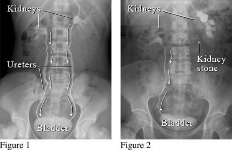
These figures show an X-ray with contrast dye (intravenous pyelogram, or IVP) of the kidneys, ureters, and bladder. Figure 1 shows a normal flow from the kidneys, through the ureters, to the bladder (white arrows). Figure 2 shows a kidney stone blocking the normal flow of urine in the ureter on the right.
Current as of: March 26, 2025
Author: Ignite Healthwise, LLC Staff
Clinical Review Board
All Ignite Healthwise, LLC education is reviewed by a team that includes physicians, nurses, advanced practitioners, registered dieticians, and other healthcare professionals.
Current as of: March 26, 2025